Dystocia in Cats: Signs, Causes, Treatment and Difficult Birth
Introduction:
What should you do if a pregnant cat appears to be in labor but a kitten is not being delivered? Dystocia in cats refers to a difficult, delayed, or abnormal labor process where a queen is unable to expel kittens from the birth canal without intervention. While feline births—often called queening—frequently proceed without human involvement, birthing complications can escalate into life-threatening emergencies for both the mother and her offspring. Understanding what constitutes abnormal labor allows cat owners and breeders to act swiftly when professional help is required.
Recognizing the signs of dystocia in cats early is critical to ensuring the health and survival of the entire litter. This comprehensive guide covers the definition of difficult labor, primary clinical signs, underlying maternal and fetal causes, emergency at-home steps, veterinary diagnosis, medical and surgical treatment options, post-labor recovery, and post-birth complications.
What Is Dystocia in Cats?
What Does Dystocia Mean?
The term dystocia is derived from the Greek roots dys- (meaning difficult, painful, or abnormal) and tokos (meaning childbirth). In feline medicine, dystocia denotes an inability to deliver fetuses through the birth canal during stage two of labor.
Feline labor is categorized into three distinct stages:
- Stage One: The queen becomes restless, seeks a quiet birthing area (nesting), exhibits temperature drops, and experiences uterine contractions without visible abdominal straining. Cervical dilation occurs during this period, which typically lasts between 6 and 12 hours (though it can extend up to 24 hours in nervous or first-time mothers).
- Stage Two: Active labor begins. Strong, visible abdominal contractions start, the amniotic sac ruptures, and kittens are expelled.
- Stage Three: Delivery of the placenta occurs, usually following each kitten or alternating with subsequent kitten deliveries.
Dystocia occurs when stage two fails to progress normally due to functional, physical, or anatomical abnormalities.
Normal Cat Labor vs. Difficult Birth

| Clinical Parameter | Normal Feline Birthing Process | Suspected Feline Dystocia |
| Active Straining | Strong abdominal contractions leading to a kitten birth within 30 minutes. | Strong, persistent contractions lasting longer than 30 minutes without delivering a kitten. |
| Rest Period Between Kittens | Mild, resting contractions or calm resting phase lasting 30 minutes to 2 hours between kittens. | Weak, infrequent, or absent contractions for over 2 hours when additional fetuses remain inside. |
| Maternal Behavior | Mother is alert, grooming kittens, and purring despite labor exertion. | Mother exhibits extreme distress, persistent crying, constant licking of the vulva, or biting at her rear. |
| Physical Condition | Mother remains stable, hydratable, and responsive. | Mother shows profound weakness, lethargy, tremors, or collapse. |
| Vaginal Discharge | Clear, mucous, or slight bloody discharge; green/dark discharge accompanying kitten delivery. | Foul-smelling, heavy bright-red bleeding, or dark green/black discharge before any kitten is born. |
Why Dystocia Requires Immediate Attention
Unlike prolonged labor in some other species, feline labor complications carry a high risk of fetal death due to placental separation. When uterine contractions detach the placenta from the uterine wall, the fetus loses its oxygen supply. If the kitten is not expelled promptly, fetal hypoxia, brain damage, and death occur rapidly. Furthermore, retained fetuses or prolonged uterine rupture can lead to severe maternal sepsis, peritonitis, hemorrhage, and death.
Signs of Dystocia in Cats
How Do I Know If My Cat Is Having Trouble Giving Birth?
Determining when feline labor has crossed from normal physiological exertion into dangerous dystocia requires careful observation. Feline labor difficulties manifest through a variety of behavioral, reproductive, and systemic signs:
- Persistent Unproductive Straining: Strong abdominal contractions occurring continuously for 30 minutes or longer without the passage of a fetus.
- Prolonged Interuterine Delay: A pause of more than 2 hours between kittens when the owner knows additional fetuses remain in the abdomen (verified by prior X-rays or abdominal palpation).
- Visible Fetus in the Birth Canal: A kitten’s tail, head, or feet visible at the vulvar opening for more than 10 to 15 minutes without progress despite maternal effort.
- Abnormal Vaginal Discharge: Passage of dark green, brown, or black discharge (uteroverdin) before the first kitten has been delivered indicates premature placental detachment. Heavy, frank red arterial blood issuing from the vulva also signifies uterine tear or vascular rupture.
- Systemic Maternal Illness: Extreme lethargy, muscle tremors, severe weakness, collapse, pale gums, vomiting, or abnormal vocalizations indicating agonizing pain.
Dystocia Signs That Require Urgent Veterinary Attention
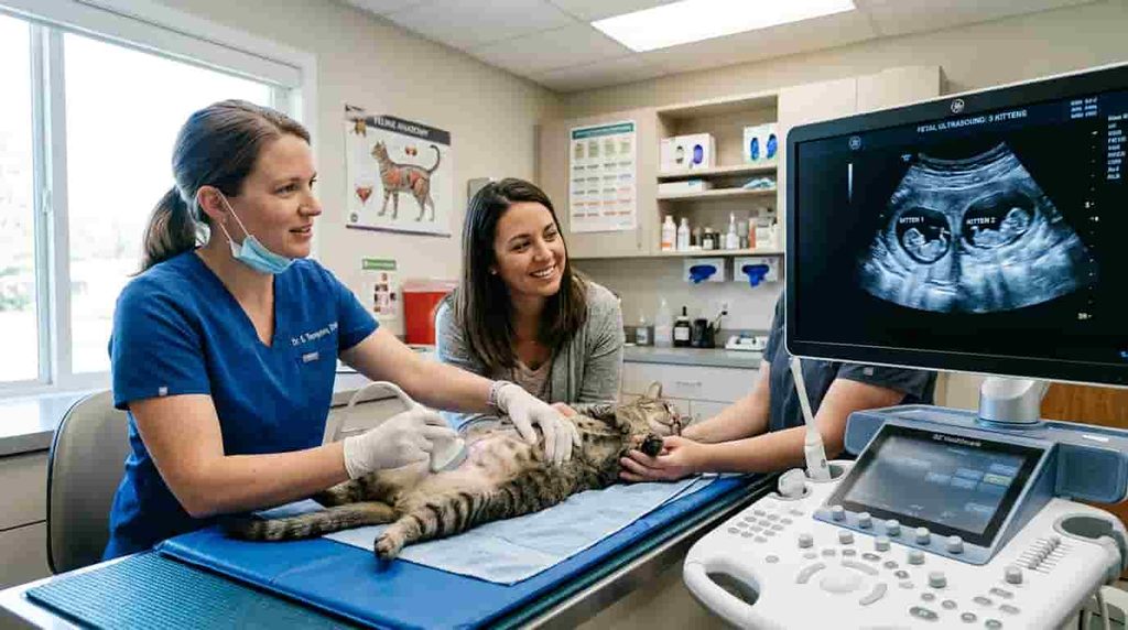
If a pregnant cat displays any of the following clinical signs, immediate transport to an emergency veterinary clinic is mandatory:
- Active straining for over 30 minutes with no kitten produced.
- More than 2 hours elapsed since the last kitten was born, with confirmed fetuses remaining.
- Greenish or purulent discharge prior to the birth of the first kitten.
- Gestation extending past 68 to 70 days from the last breeding date.
- Presence of a stuck fetus or membranes extending from the vulva that cannot be safely dislodged.
- Maternal exhaustion, fever, hypothermia, or loss of consciousness.
What Owners Should Observe and Record
When monitoring a birthing queen, keep a precise log to assist your veterinarian during assessment:
- Timeline: Record when stage one labor behaviors started (nesting, temp drop) and the exact time active stage two straining commenced.
- Delivery Gaps: Note the birth time of each kitten and whether a corresponding placenta was expelled.
- Physical Effort: Document whether contractions are visible, strong, or intermittent.
- Discharge Characteristics: Track the color, volume, and timing of any vulvar discharge relative to kitten deliveries.
What Causes Dystocia in Cats?
Dystocia is broadly classified into two pathophysiological mechanisms: maternal causes (originating from the mother) and fetal causes (originating from the kittens). It is further categorized into functional or mechanical origins.
Maternal Causes
Maternal factors account for approximately 60% to 70% of feline dystocia cases.
1. Uterine Inertia (Primary vs. Secondary)
Uterine inertia is the failure of the uterine smooth muscle (myometrium) to contract forcefully enough to expel a fetus.
- Primary Uterine Inertia: The uterine muscle fails to initiate functional contractions. Causes include hormonal imbalances (progesterone non-decline), small litter sizes (one or two kittens failing to produce sufficient hormonal triggers for labor), extremely large litters causing overstretching of the myometrium, hypocalcemia (low blood calcium), or systemic maternal disease.
- Secondary Uterine Inertia: The uterine muscle contracts normally at first but eventually becomes exhausted following prolonged, unsuccessful attempts to clear an obstructed birth canal.
2. Anatomical Obstructions
Physical narrowing of the birth canal prevents passage of normal-sized fetuses:
- Pelvic Canal Deformities: Previous pelvic fractures (often from motor vehicle trauma) that healed abnormally reduce the pelvic diameter.
- Soft Tissue Abnormalities: Vaginal strictures, congenital abnormalities, uterine torsion (twisting of a uterine horn), uterine rupture, or vaginal tumors.
- Cervical Malfunction: Inability of the cervix to fully dilate during stage one labor.
Fetal Causes
Fetal factors account for 30% to 40% of feline dystocia cases.
1. Abnormal Presentation, Posture, and Position
While anterior presentation (head-first with legs extended) and posterior presentation (tail and hind legs first—often called breech) are both considered normal in cats, abnormal orientations cause mechanical blockage:
- Transverse Presentation: The fetus lies sideways across the opening of the birth canal.
- Head Inversion or Flexion: The head is bent downward or backward, presenting the nape of the neck rather than the snout to the pelvis.
- Limb Flexion: One or both front limbs are bent backward beneath the body, increasing the cross-sectional width of the fetus.
2. Fetal Oversize (Relative or Absolute)
- Single-Kitten Litters: When only one fetus develops, it receives an abundant nutrient supply in utero, leading to exaggerated size (fetal gigantism) that exceeds the diameter of the mother’s pelvic inlet.
- Fetal Malformations: Congenital anomalies such as hydrocephalus (fluid accumulation on the brain), anasarca (severe fetal subcutaneous edema), or joined twins prevent passage through the pelvic ring.
- Fetal Death: A dead fetus cannot align itself actively with the birth canal and lacks normal fluid pressure mechanics, predisposing labor to failure.
Mechanical vs. Functional Dystocia
- Mechanical Dystocia: Occurs when a physical barrier (oversized fetus, abnormal presentation, narrow pelvis) stops the passage of a kitten. Uterine contractions may be vigorous initially.
- Functional Dystocia: Occurs when the birth canal is unobstructed, but the uterine muscles fail to contract effectively due to metabolic, muscular, or hormonal deficiencies.
Risk Factors for Cat Pregnancy Problems

Certain breeds, health conditions, and environmental factors predispose pregnant cats to birthing complications.
Factors That May Increase Concern
- Breed Predispositions: Brachycephalic breeds (such as Persians, Exotic Shorthairs, and Himalayans) have broader, flatter head structures that increase the risk of fetal-pelvic disproportion. Purebred cats generally have a higher incidence of dystocia compared to mixed-breed domestic shorthairs.
- Maternal Age: Extremely young queens (under 1 year of age) whose pelvic bones have not fully fused, or elderly queens (over 6–7 years) with declining uterine muscle tone, face higher complication rates.
- History of Prior Dystocia: A queen that has experienced a difficult birth in a previous litter is significantly more likely to encounter dystocia in subsequent pregnancies.
- Pre-existing Trauma: Any history of pelvic trauma, fractures, or abdominal surgery.
- Nutritional and Metabolic Disturbances: Malnutrition, obesity, or improperly balanced diets leading to gestational hypocalcemia or hypoglycemia during labor.
Why Pregnancy Monitoring Matters
Proactive monitoring during the 63 to 65 days of feline gestation reduces emergency risks. Abdominal radiography (X-rays) performed by a veterinarian during the last week of pregnancy (after day 55, when fetal skeletons calcify) allows for precise fetal counts and evaluation of head-to-pelvis ratios. Ultrasound imaging assesses fetal heart rates and viability.
Simple Preparation Checklist
Before the estimated queen delivery date:
- Confirm the expected litter size with a late-term veterinary X-ray.
- Identify an accessible 24-hour emergency veterinary clinic and save their contact details.
- Assemble a birthing kit: clean towels, blunt-nosed sterile scissors, unwaxed dental floss (for tying umbilical cords), warm water bottles, and an infant suction bulb.
- Prepare a quiet, warm, draft-free nesting box in an isolated room.
- Record baseline rectal temperatures twice daily during the final week (a drop below 99°F / 37.2°C often signals labor within 12–24 hours).
What Should You Do If You Suspect Dystocia?

How to Help a Cat with Dystocia
If you suspect your cat is experiencing difficult labor, the most crucial action is to immediately contact a qualified veterinarian or emergency animal hospital. Dystocia in cats is a medical emergency that cannot be safely managed at home with home remedies or manual traction. Attempting to manage feline labor failure without professional diagnostic equipment frequently leads to kitten death, uterine rupture, or fatal maternal infection.
What You Can Safely Do While Seeking Veterinary Care
- Remain Calm: Feline queens are acutely sensitive to owner stress. High environmental stress causes adrenaline release, which actively inhibits oxytocin release and slows uterine contractions.
- Keep the Mother Warm and Quiet: Place her in a dimly lit, comfortable carrier lined with dry towels.
- Minimize Movement: Avoid squeezing her abdomen or placing pressure on her pelvic region.
- Call Ahead: Contact the emergency clinic so the veterinary medical team can prepare an examination room, warm incubator, diagnostic imaging, and an operating suite for a potential emergency C-section.
- Transport Safely: Maintain a warm vehicle interior during transport, as hypothermia rapidly destabilizes both mother and newborn kittens.
What NOT to Do
- DO NOT pull forcefully on exposed fetal limbs or tail. Pulling on a stuck fetus without proper lubrication and veterinary expertise can cause traumatic limb amputation, pelvic fracture in the mother, or fatal uterine rupture.
- DO NOT administer human or non-prescribed medications. Drugs meant to stimulate labor (like oxytocin) can tear the uterus if administered during a mechanical obstruction.
- DO NOT perform internal digital examinations. Inserting unsterile fingers into the birth canal risks introducing pathogenic bacteria directly into an open, vulnerable uterine cavity.
- DO NOT wait overnight or “see if she passes it by morning.” Delays exceeding a few hours drastically reduce kitten survival rates and compromise maternal life.
Veterinary Diagnosis of Dystocia
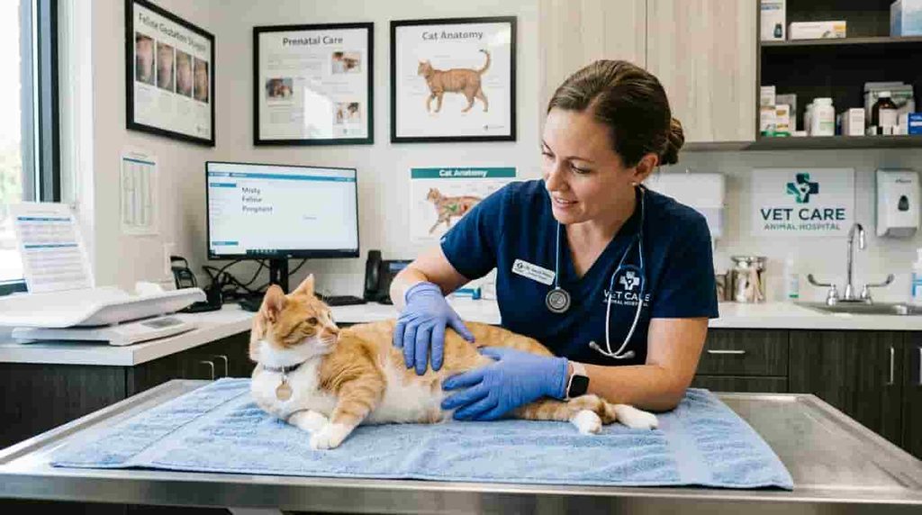
When a queen arrives at a veterinary hospital for suspected dystocia, the clinical team performs a rapid, systematic evaluation to determine whether medical management or emergency surgical intervention is indicated.
Physical Examination
The veterinarian will conduct a targeted triage exam:
- Systemic Assessment: Evaluation of maternal heart rate, gum color, capillary refill time, hydration status, body temperature, and blood pressure.
- Abdominal Palpation: Checking for remaining fetuses, uterine muscle tone, and signs of abdominal pain or uterine rupture.
- Digital Vaginal Examination: Assessment of vaginal dilation, presence of strictures, pelvis dimensions, and identification of any fetus positioned within the pelvic inlet. The Ferguson reflex (feathering the dorsal vaginal wall to stimulate uterine contractions) may also be evaluated.
Imaging and Diagnostic Testing
- Abdominal Radiographs (X-rays): Crucial for determining the exact number, size, and location of remaining fetuses, evaluating fetal position/presentation, and checking for fetal death (indicated by gas formation in fetal tissues or overlapping cranial bones).
- Ultrasonography: Essential for evaluating fetal viability and distress. A normal fetal feline heart rate ranges between 180 and 220 beats per minute (bpm). A fetal heart rate dropping below 160 bpm indicates fetal distress, while rates under 140 bpm demand immediate emergency surgical extraction to prevent fetal mortality.
- Blood Work: Diagnostic blood panels evaluate maternal packed cell volume (PCV), blood glucose levels, and serum ionic calcium concentration. Hypocalcemia and hypoglycemia are common metabolic triggers for functional uterine inertia.
Determining the Cause
By correlating clinical findings, blood work, radiographs, and ultrasound heart rates, the veterinarian determines whether the dystocia is functional or mechanical, allowing them to select the safest treatment path.
Dystocia in Cats Treatment
Treatment strategies range from conservative medical therapy to emergency surgical delivery (cesarean section), depending directly on the underlying diagnostic findings.
What Is the Treatment for Dystocia?
Veterinary management aims to safely deliver all fetuses while preserving the health of the queen:
- Conservative / Manual Assistance: If a fetus is lodged in the vagina in a normal presentation and no physical obstruction exists, a veterinarian may apply sterile lubricant and gently guide the fetus during active maternal contractions using specialized techniques.
- Medical Management: Indicated strictly when there is no mechanical obstruction present, the mother is in good systemic health, the cervix is dilated, and fetal heart rates indicate no severe distress.
- Surgical Intervention (C-Section): Indicated when mechanical obstruction exists, medical treatment fails, fetal heart rates reveal distress, or uterine rupture/torsion is present.
What Medications Are Used to Treat Dystocia?
Medical intervention must always be supervised directly by a veterinarian following diagnostic imaging:
- Oxytocin: A hormone that stimulates smooth muscle contractions in the myometrium. It is administered in low, precise micro-doses intramuscularly or intravenously. Oxytocin is strictly contraindicated if a mechanical obstruction exists, as strong contractions against a blocked birth canal can cause fatal uterine rupture.
- Calcium Gluconate: Administered slowly intravenously or subcutaneously to improve myometrium contraction strength, especially if blood tests confirm low calcium levels. Calcium administration sensitizes the uterine muscle to the action of oxytocin.
- Dextrose Solution: Administered intravenously to correct maternal hypoglycemia and provide metabolic energy for labor effort.
If oxytocin and calcium administration fail to produce a kitten delivery within 30 to 40 minutes, medical therapy is deemed unsuccessful, and immediate surgery is initiated.
When Is a C-Section Needed?
A cesarean section (hysterotomy) is required in the following circumstances:
- Confirmed Mechanical Obstruction: Fetal oversize, pelvic narrowing, or birth canal deformities.
- Fetal Distress: Fetal heart rates consistently below 150–160 bpm on ultrasound.
- Uterine Inertia Unresponsive to Medication: Failure of oxytocin and calcium to induce productive labor.
- Anatomical Accidents: Uterine torsion, rupture, or severe systemic maternal collapse.
- Dead or Decomposing Fetuses: Presence of dead fetuses obstructing the passage.
During a C-section, the queen is placed under general anesthesia, an incision is made along the abdominal midline, the uterine horns are exteriorized, and fetuses are gently extracted. An ovariohysterectomy (spaying) may be performed concurrently if the uterine wall is necrotic, infected, or if the owner chooses not to breed the queen again.
Recovery After a Difficult Cat Birth
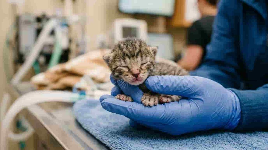
Post-operative care and recovery following dystocia management require diligent home monitoring.
Caring for the Mother After Treatment
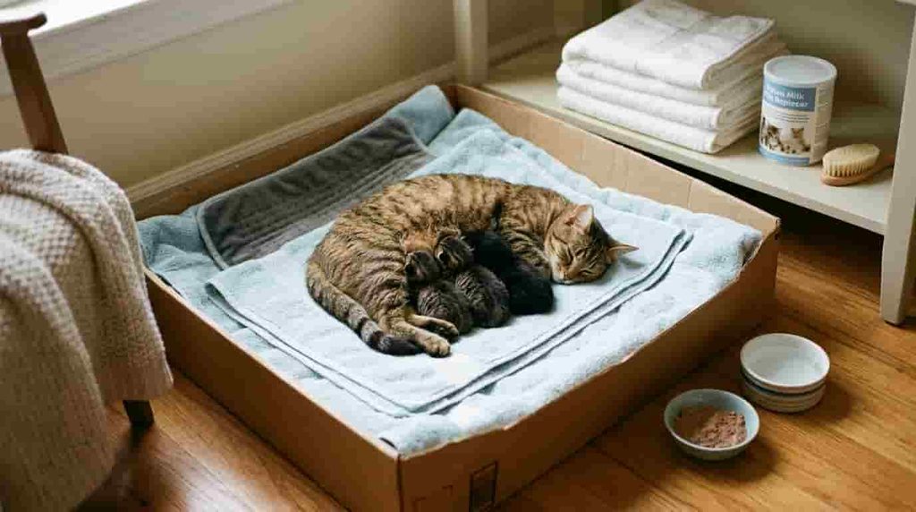
- Environment: Provide a quiet, warm, isolated nursing sanctuary away from household noise and other pets.
- Pain Management and Medications: Administer all veterinarian-prescribed analgesics, antibiotics, or anti-inflammatory drugs precisely as directed. Ensure any prescribed pain medication is strictly feline-safe and safe for nursing kittens.
- Incisions and Hygiene: Inspect abdominal surgical incisions daily for redness, swelling, discharge, or missing sutures. Gently clean the vulvar area if lochia (normal post-birth discharge) accumulates.
Monitoring the Kittens
- Warmth and Feeding: Newborns cannot regulate their own body temperature. Ensure they nurse successfully within 2 to 4 hours of birth to absorb crucial colostrum antibodies. If the mother is recovering from general anesthesia, supplemental bottle feeding with a commercial feline milk replacer may be required initially.
- Daily Weight Gain: Track newborn weights daily using a digital gram scale. Healthy kittens should gain 10 to 15 grams per day after the first 24 hours.
When to Contact the Veterinarian Again
Contact your veterinary hospital immediately if:
- The queen refuses to eat, develops a fever (temperature >102.5°F / 39.2°C), or exhibits profound lethargy.
- The surgical incision gapes, becomes red, or leaks foul-smelling fluid.
- The queen rejects her kittens or shows aggressive behavior toward them.
- Kittens cry continuously, lose weight, or become cold and unresponsive.
Possible Complications After Dystocia
Even after all kittens are delivered, queens recovering from difficult labor face risks of secondary reproductive and systemic complications.
Why Monitoring Does Not End After Delivery
Retained placental tissue, uterine trauma, or bacterial contamination during prolonged labor increases susceptibility to post-partum infections. Watch closely for heavy, foul-smelling vaginal discharge, persistent vomiting, or systemic illness during the first 2 to 3 weeks post-partum.
Connection to Mastitis
Dystocia and systemic maternal stress can impair milk flow or lead to ascending bacterial infections in the mammary glands. A queen experiencing swelling, heat, pain, or discoloration in her nipples may be developing mastitis. For detailed information on identifying and treating swollen or infected milk glands, refer to our complete guide on Mastitis in Cats: Symptoms, Causes, Treatment and When to See a Vet
Connection to Pyometra
Bacterial invasion of the uterus during a prolonged, obstructed labor can progress to severe uterine infection. While pyometra typically occurs during the luteal phase of the estrous cycle, post-parturient uterine infections (metritis) present with similar life-threatening septic symptoms and vaginal discharge. To learn more about serious uterine infections, diagnosis, and emergency surgical options, read our article on Pyometra in Cats: Symptoms, Causes, Treatment, Surgery and Survival
Can Dystocia in Cats Be Prevented?

While not all causes of dystocia can be eliminated, proactive breeding management significantly reduces overall incidence rates.
Responsible Pregnancy Care
- Pre-Breeding Health Screening: Avoid breeding queens with known histories of pelvic trauma, structural abnormalities, or prior labor failure. Ensure both breeding parents undergo genetic and physical evaluations.
- Optimal Nutrition: Feed a high-quality, balanced growth/lactation formula during the final third of pregnancy. Avoid overfeeding, as maternal obesity directly narrows the pelvic birth canal with soft tissue fat deposits. Avoid unverified calcium supplementation during pregnancy, as it paradoxically disrupts normal parathyroid gland function and increases the risk of labor inertia.
- Veterinary Oversight: Schedule late-gestation diagnostic imaging to confirm fetal counts and evaluate pelvic adequacy.
Know the Cat’s Reproductive History
If a queen requires an emergency C-section due to anatomical pelvic stenosis or primary uterine inertia, she should be spayed to prevent future life-threatening labor complications.
Dystocia Management: A Practical Emergency Checklist
Before Labor
- Emergency clinic contact details and location saved.
- Transport carrier equipped with dry, warm towels.
- Late-term diagnostic X-ray completed to confirm fetal numbers.
- Quiet, draft-free nesting box prepared.
During Labor
- Record the exact time active stage two straining starts.
- Note intervals between each kitten birth.
- Monitor the mother continuously for weakness or intense distress.
- Inspect discharge color (green or dark discharge before the first kitten requires an emergency visit).
If Dystocia Is Suspected
- Contact an emergency veterinary clinic immediately.
- Do not pull forcefully on stuck fetuses or fetal tissue.
- Do not administer unprescribed medications or home remedies.
- Transport the queen carefully in a temperature-regulated vehicle.
Dystocia in Cats vs. Dystocia in Other Animals
Why Dystocia Is Not Managed the Same Way in Every Animal
While the term dystocia is used across veterinary medicine to describe birthing difficulties in dogs, cattle, horses, and sheep, the diagnostic criteria, physiological limits, and treatment approaches vary significantly by species:
- Canine vs. Feline Dystocia: Dogs experience a higher rate of obstructive dystocia due to dramatic skull size variations among breeds (e.g., Bulldogs). In contrast, feline dystocia is more frequently related to uterine inertia or malpositioned fetuses. Medical dosage protocols and surgical techniques also differ substantially between species.
- Large Animal Dystocia: Livestock management (such as cattle or sheep) often utilizes manual traction devices (calving chains) and fetotomy procedures. These methods are catastrophically dangerous if applied to cats due to the fragile nature of feline tissues and the small size of the queen’s birth canal.
Always rely on feline-specific veterinary medical standards when addressing pregnancy complications in cats.
When Is Cat Labor an Emergency?
Seek immediate emergency veterinary care if you observe any of the following critical indicators:
- More than 30 minutes of continuous, strong abdominal straining without delivering a kitten.
- More than 2 hours between kittens with known fetuses remaining.
- Appearance of green, black, or heavy bloody vulvar discharge prior to the birth of the first kitten.
- A fetus visible in the birth canal that is not expelled within 10 to 15 minutes.
- Maternal exhaustion, collapse, muscle tremors, or pale gums.
- Pregnancy lasting longer than 68 days from confirmed mating.
Emergency Rule: When in doubt regarding the progress of feline labor, consult a veterinarian immediately. Early intervention preserves lives; waiting rarely improves the outcome of an obstructed labor.
Frequently Asked Questions
FAQ 1: How do I know if my cat is having trouble giving birth?
Signs of labor trouble include strong abdominal straining for over 30 minutes without producing a kitten, gaps exceeding 2 hours between kitten deliveries, green or bloody discharge before the first birth, or maternal collapse. If your cat displays severe distress or lethargy, contact a veterinarian immediately.
FAQ 2: What is the treatment for dystocia?
Treatment depends on the underlying cause determined by veterinary examination and X-rays. Options range from manual assisted delivery and drug therapy (such as oxytocin and calcium) to emergency surgical delivery via a C-section.
FAQ 3: What medications are used to treat dystocia?
Veterinarians primarily use oxytocin to stimulate uterine contractions, along with calcium gluconate and dextrose to support muscle performance and energy. These medications are strictly administered under professional guidance when no physical obstruction is present.
FAQ 4: How can I help a cat with dystocia?
The safest way to help is to keep the mother calm, warm, and quiet while immediately transporting her to an emergency veterinary clinic. Never pull forcefully on exposed fetuses or administer home remedies, as this can cause fatal injury.
FAQ 5: Can dystocia happen again in cats?
Yes, a queen that has experienced dystocia is at high risk for recurring labor difficulties in future pregnancies. If the underlying cause was anatomical pelvic narrowing or uterine inertia, spaying the cat is strongly recommended.
Conclusion
Recognizing dystocia in cats quickly can mean the difference between life and death for both the mother and her litter. Whether caused by primary uterine inertia, fetal oversize, or abnormal presentation, difficult labor requires prompt diagnostic assessment by a qualified veterinarian. By monitoring your cat’s pregnancy, establishing an emergency plan ahead of time, and seeking immediate professional assistance when warning signs appear, you ensure the highest standard of care for your feline companion.
Have you experienced a difficult birthing process with your pregnant cat, or do you have questions about preparing your nesting area? Share your experiences or ask your questions in the comments below!